
Attorney's Guide to Radiology Imaging
Everything attorneys need to know about radiology: imaging modalities, reading reports, ambiguous language, second opinions, visual documentation, and the opposing-counsel playbook.

Legal disclaimer
Educational material only. This guide is not legal advice, does not create an attorney-client relationship, and does not substitute for counsel licensed in the relevant jurisdiction.
Table of Contents
- 1. The Imaging Timeline: From Accident to Demand Letter
- 2. MRI, CT, X-ray, and Ultrasound: What Each One Shows
- 3. How to Read a Radiology Report
- 4. The Ambiguous Language Problem
- 5. Five Mistakes Attorneys Make with Imaging Evidence
- 6. The Value of Second Opinions
- 7. Visual Evidence and Jury Comprehension
- 8. The Opposing Counsel Playbook: What the Other Side Will Do
- 9. Expert Witness Credibility
Key Takeaways
- • Objective imaging findings are harder to dispute than subjective descriptions alone. Early advanced imaging can create a clearer evidentiary timeline.
- • MRI is the gold standard for many soft tissue injuries. Public settlement benchmarks vary widely and should not be treated as outcome guarantees.
- • Phrases like "may be consistent with" and "cannot exclude" can create avoidable ambiguity that legal teams need to evaluate carefully.
- • Subspecialty second opinions reveal a 32.2% discrepancy rate with original reads, and 20.4% of those discrepancies are major enough to change case management.
- • Visual evidence can improve comprehension. Illustrated reports and medical animations should be framed as explanation tools, not outcome guarantees.
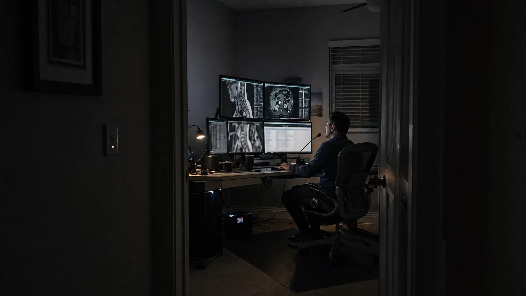
1. The Imaging Timeline: From Accident to Demand Letter
Every injury case has an imaging timeline, and the clarity of the record often depends on how well that timeline was managed. Imaging evidence is not just supporting documentation. It can become the objective foundation for demand materials, mediation presentations, and trial strategy.
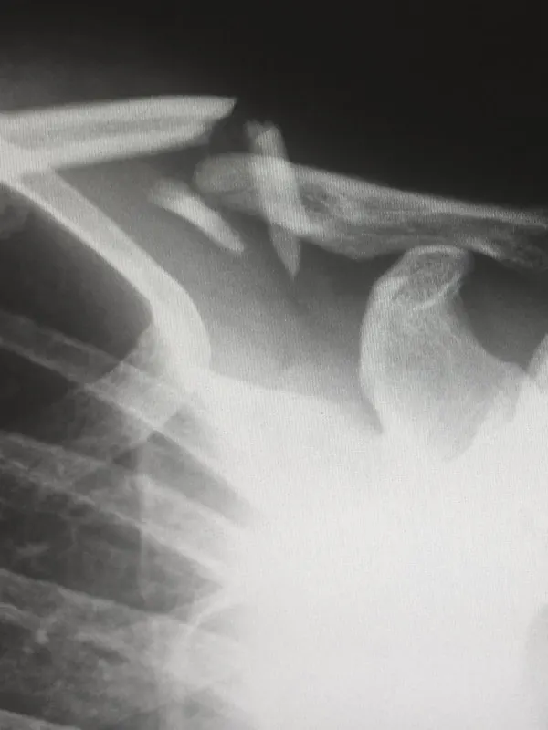
The sequence typically follows a predictable path. The patient arrives at the emergency room, where X-rays are ordered first to rule out fractures, dislocations, and other acute skeletal injuries. X-rays are fast, cheap, and widely available. They are also severely limited. They show bones and gross alignment, but they cannot visualize the soft tissue injuries that drive many injury claims: disc herniations, ligament tears, labral pathology, rotator cuff damage, and nerve compression.
Advanced imaging may be useful early when clinically appropriate. MRI and CT scans performed close in time to the incident can create a contemporaneous record. Timing matters because reviewers may scrutinize gaps between the date of injury and the date of imaging.
Once imaging is obtained, interpretation happens on two different timelines. STAT reads for emergency and urgent cases typically come back within one to two hours. Routine interpretations take approximately 24 hours. The turnaround time matters because the treating physician needs findings to guide the treatment plan, and the treatment plan itself becomes evidence.
Here is the chain: accident leads to ER evaluation (X-rays), which leads to advanced imaging (MRI or CT), which leads to radiology interpretation, which guides treatment based on findings, which ultimately feeds the demand letter. At every link in this chain, the imaging evidence either strengthens or weakens your position. Your job is to make sure every link holds.
2. MRI, CT, X-ray, and Ultrasound: What Each One Shows
Choosing the wrong imaging modality is one of the fastest ways to weaken a case before it even gets started. Each modality has specific strengths and specific blind spots. Understanding what each one actually shows (and what it misses) is not optional for attorneys handling imaging-heavy injury claims.
X-ray ($100 to $300). X-rays are the most basic imaging modality. They excel at showing fractures, dislocations, and gross skeletal alignment. Results are available same-day, often within minutes. The limitation is significant: X-rays cannot visualize soft tissue structures in any meaningful detail. If your client's injury involves a disc herniation, ligament tear, or nerve compression, an X-ray will show nothing. A clean X-ray does not mean a clean bill of health.
CT scan ($300 to $3,280). CT provides cross-sectional images using X-ray technology. It is the modality of choice for complex fractures, acute hemorrhage, and detailed bone anatomy. CT is faster than MRI and handles acute trauma cases well. The critical limitation: CT frequently misses soft tissue injuries. ACL tears, rotator cuff damage, meniscal pathology, and disc herniations can all be overlooked on CT. According to Johns Hopkins, CT and MRI serve fundamentally different diagnostic purposes, and using CT when MRI is indicated means missing the findings that matter most in litigation.
MRI ($400 to $12,000; average $1,325). MRI is the gold standard for many soft tissue evaluations. It visualizes disc herniations, ligament tears, tendon pathology, cartilage damage, bone marrow edema, and nerve compression with a level of detail no other modality can match. For injury cases, MRI is often the definitive imaging study when soft tissue injury is central to the claim. Public legal-market sources report wide settlement differences when structural pathology is documented, but those figures are case-specific and not outcome guarantees. Our MRI vs. CT guide covers the clinical decision-making in greater detail.
Ultrasound ($150 to $592). Ultrasound uses sound waves to create real-time images. It is best for superficial soft tissue evaluation: fluid collections, superficial tendon tears, and guided injections. It is portable, inexpensive, and does not use radiation. However, ultrasound cannot penetrate deep structures and is highly operator-dependent. It has a role in certain MSK assessments but rarely serves as primary litigation evidence for major injury claims.
The bottom line: if a claim depends on demonstrating soft tissue injury, MRI is often the study that answers the question. Relying on X-rays alone because they were already obtained in the ER can leave the record incomplete.
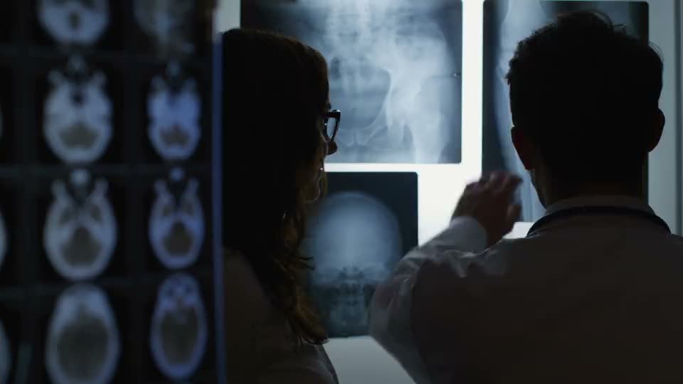
3. How to Read a Radiology Report
A radiology report is not a mystery document. It follows a standardized structure established by the American College of Radiology and used by virtually every radiology practice in the country. Once you understand the five sections and what to look for in each, you can evaluate any report in minutes.
1. Clinical Indication. This section states why the study was ordered. It includes the patient's symptoms, the mechanism of injury, and the clinical question the referring physician wants answered. Pay attention here: if the indication says "low back pain, rule out disc herniation" but the radiologist never specifically addresses disc pathology in the findings, that is a gap you need to flag.
2. Technique. This describes how the study was performed: the sequences used, whether contrast was administered, the plane of imaging. Technique matters because certain findings are only visible on specific sequences. For example, a STIR sequence highlights bone marrow edema (a sign of acute injury), while standard T1-weighted images do not. If a critical sequence was omitted, it may explain why a finding was missed.
3. Comparison. This tells you whether the radiologist had prior imaging studies available for comparison. Comparison studies are enormously valuable because they allow the reader to distinguish new, acute findings from pre-existing conditions. If the comparison line says "None available," consider whether obtaining prior studies could strengthen or clarify the case.
4. Findings. This is the body of the report. The radiologist describes every anatomical structure evaluated and every abnormality identified, level by level, structure by structure. This section is typically the longest and most detailed. For spine MRIs, findings are organized by vertebral level (L3-L4, L4-L5, L5-S1, and so on). For joint studies, findings are organized by anatomical structure (ligaments, tendons, cartilage, bones).
5. Impression. This is the most important section. The impression is the radiologist's summary and synthesis of the findings. It lists the primary diagnoses, ranks them by clinical significance, and may include recommendations for further evaluation. The impression is what referring physicians read first and what opposing counsel will scrutinize most closely. A weak or ambiguous impression undermines the entire study.
What attorneys should specifically look for: measurements (a 6mm disc herniation is different from "a small herniation"), laterality (left versus right, documented consistently), acuity indicators (acute bone marrow edema versus chronic degenerative changes), comparison with prior imaging when available, and follow-up recommendations. Every one of these details either supports or weakens your case narrative. Read the impression first, but always go back to the findings for the specifics.
4. The Ambiguous Language Problem
Radiology reports should communicate supported findings clearly while preserving uncertainty when the imaging genuinely requires it. The problem is not appropriate caution; it is unnecessary ambiguity that makes the record harder to interpret.
These are the specific phrases that legal teams should review carefully:
"May be consistent with." This phrase may be appropriate in some contexts, but it can also leave reviewers unsure whether the finding is actually supported by the images.
"Cannot exclude." This phrase often signals that the reader cannot fully characterize the finding. A second read may help clarify whether the images support a more specific interpretation.
"Correlate clinically." This phrase appears in thousands of radiology reports every day, and according to Kuzminski's analysis in the Journal of the American College of Radiology (2015), it can be functionally vague. In legal review, it may create a gap between the imaging finding and the clinical record.
"Degenerative changes." As the attorneys at Miller & Zois have described it, this phrase is "liquid courage for adjusters." When a radiologist attributes findings to "degenerative changes" without distinguishing between pre-existing degeneration and acute traumatic injury superimposed on degeneration, the other side uses it to argue that the entire condition predates the accident. That distinction between chronic degeneration and acute injury is the single most important characterization in an injury-case radiology report.
The research confirms this is not a minor issue. A 2016 study published in PubMed (PMID: 27657356) found that 11 of 36 commonly used radiology terms were interpreted differently by referring clinicians than by radiologists. Phrases that radiologists considered routine were understood to mean something entirely different by the physicians acting on them. In one striking example, the study compared follow-up ordering behavior: when a report described a "benign cyst," only 2% of providers ordered follow-up imaging. When the same finding was described as "most likely cyst, but tumor cannot be excluded," 75% ordered follow-up. Same imaging. Same finding. Radically different clinical response based entirely on word choice.
At larger scale, Bhatnagar et al. analyzed 642,569 report impressions across multiple institutions and documented significant variability in language use across radiologists interpreting identical pathology. The study, published in PMC in 2020, confirmed what practicing attorneys already know: two radiologists looking at the same images can produce reports with dramatically different language, and that language directly impacts clinical decision-making.
The takeaway is simple. Ambiguous language should be reviewed, not ignored. If the report does not clearly explain the imaging basis and confidence level, a subspecialty second opinion may help clarify the record.
5. Five Mistakes Attorneys Make with Imaging Evidence
After reviewing thousands of injury-case imaging reviews, we see the same five mistakes repeatedly. Each one is avoidable.
Mistake 1: Ordering imaging too late. Insurance companies and opposing attorneys track the gap between the date of injury and the date of first advanced imaging down to the day. A gap of two weeks or more is an invitation for the argument that the injury was not severe enough to warrant urgent evaluation, or that something else caused it. When your client's treating physician recommends imaging, make sure it happens immediately. Follow up. A delay that seems insignificant clinically can be devastating in litigation.
Mistake 2: Using the wrong modality. An X-ray cannot show a disc herniation. A CT scan routinely misses ligament tears. If your client's injury is soft tissue based, ordering only X-rays or relying solely on the ER's initial CT is insufficient. Make sure the treating physician orders the modality that actually visualizes the injury. For soft tissue, that means MRI. See our detailed MRI vs. CT comparison for modality selection guidance.
Mistake 3: Not getting a second opinion. According to the meta-analysis by Rosenkrantz et al., published in the Journal of the American College of Radiology in 2018, the overall discrepancy rate between initial radiology reads and subspecialty second opinions is 32.2%. More than one in three original reads contains findings that a subspecialist would characterize differently. Of those discrepancies, 20.4% are classified as major, and 18.6% are significant enough to change clinical management. If you accept the initial read at face value without considering a second opinion, you are rolling the dice on evidence that has a one-in-three chance of being incomplete or inaccurate. Our second opinions article goes deeper on this topic.
Mistake 4: Accepting ambiguous language without review. Too many attorneys receive a report, read the impression, and move forward without evaluating the quality of the language. If the report does not clearly state what the imaging supports, a subspecialty re-read can help clarify the finding and confidence level.
Mistake 5: Not using visual evidence. A radiology report is a text document. Judges and juries are not trained to read medical text. They process visual information. According to the Weiss-McGrath study on courtroom evidence retention, jurors retain only 10% of information delivered orally. When visual evidence is combined with oral testimony, retention jumps to 65%. That is a 650% increase in evidentiary impact. If you are presenting imaging findings using only the written report and oral testimony from a radiologist, you are leaving the majority of your persuasive potential on the table.
6. The Value of Second Opinions
The clinical and documentation value of second opinions in radiology is backed by some of the most robust data in diagnostic medicine.
The landmark meta-analysis by Rosenkrantz et al. (JACR, 2018) synthesized data from dozens of studies examining the discrepancy rate between initial radiology interpretations and subspecialty second reads. The headline numbers: 32.2% overall discrepancy rate, 20.4% major discrepancy rate, and 18.6% of discrepancies significant enough to change clinical management. In practical terms, roughly one in five second opinions identifies a finding serious enough to alter the treatment plan, the diagnosis, or both.
The rates vary by modality and subspecialty. CT studies show a 28.3% discrepancy rate. MRI studies come in at 31.2%. Musculoskeletal imaging carries a 34% discrepancy rate. Body imaging (abdominal and pelvic studies) has the highest rate at 43.8%. These are not edge cases. These are core imaging modalities interpreted in high volumes every day at hospitals and imaging centers across the country.
The accuracy of second opinion reads is also well-documented. When a subspecialty second opinion disagrees with the original read, the second opinion is correct approximately 90.5% of the time. Chalian et al. (AJR, 2016) specifically examined MSK imaging and found that fellowship-trained MSK subspecialists were correct 82% of the time when their interpretation differed from a general radiologist's. A separate analysis by Chalian found a 26.2% clinically significant discrepancy rate in MSK second opinions overall.
For legal teams, the practical value is clearer documentation. If a second opinion identifies a missed disc herniation, clarifies a possible tear, or provides more precise characterization, the team has a better record to evaluate. Financial impact is matter-specific, so public tools should model assumptions rather than promise outcome lift.
Error rates in radiology are a documented reality. Brady (PMC, 2017) reviewed the literature on radiology error and found that even among experienced radiologists, perceptual errors (failing to see a finding that is present on the images) and cognitive errors (seeing a finding but misinterpreting it) are persistent. 71% of radiologists have been sued at least once during their careers. The question is not whether errors exist but whether you are catching them before they compromise your client's case.
For attorneys handling any case where imaging is central to the claim, a subspecialty second opinion can be a practical documentation safeguard. Use our Legal Imaging Value Calculator to model review-time, exhibit-prep, and report-cost assumptions for your practice.
7. Visual Evidence and Jury Comprehension
The gap between what a radiologist sees on an MRI and what a juror understands is enormous. Radiologists spend years training to interpret grayscale images full of anatomical detail. Jurors have no training at all. If you present imaging evidence as raw MRI slices and a written report, you are asking twelve untrained people to do a radiologist's job. They will not do it well.
The Weiss-McGrath research on courtroom evidence retention established the baseline: jurors retain approximately 10% of information delivered through oral testimony alone. When visual evidence is presented alongside oral testimony, retention increases to 65%. That is not a marginal improvement. That is a 650% increase in the amount of information jurors carry into deliberation. The information they retain is the information that drives verdicts.
More recent research has extended these findings. Errickson et al. published a study in the International Journal of Legal Medicine (2020) examining juror comprehension of different visual evidence formats. 3D anatomical models produced 98% juror comprehension rates. Medical animations achieved 88% comprehension. Both formats dramatically outperformed traditional 2D images and text-based evidence presentations.
Case-study sources report examples where visual exhibits were used in matters with significant outcomes. Those examples are useful for understanding how visuals may support comprehension, but they are not guarantees and should not be generalized across matters.
There is a second mechanism at work beyond jury comprehension. Visual documentation can also improve patient understanding. Treatment non-adherence rates for musculoskeletal conditions range from 26% to 63%, and clearer explanations may help patients understand what is happening inside their body and why treatment matters.
Colorized key images, custom medical illustrations, and plain-language summaries are not presentation extras. They are practical communication tools that can improve comprehension for patients, providers, and legal reviewers.
8. The Opposing Counsel Playbook: What the Other Side Will Do
If you know the playbook, you can counter every play before it develops. Opposing counsel and insurance carriers use a remarkably consistent set of strategies to undermine imaging evidence. Here are the seven moves you will see most frequently.
Play 1: "Degenerative changes." This is the other side's favorite word. If the radiologist's report mentions degenerative disc disease, degenerative joint disease, or degenerative changes of any kind, the other side will argue that the findings predate the accident. The strategy is simple: attribute the pathology to age, wear, and time rather than trauma. The counter is equally straightforward. The eggshell skull doctrine holds that a defendant takes the plaintiff as they find them. A pre-existing degenerative condition that was asymptomatic before the accident and symptomatic after it was aggravated by the accident. The key is having a radiologist who distinguishes between chronic degenerative findings and acute traumatic findings superimposed on degeneration, with specific imaging markers to support that distinction.
Play 2: The Opposing IME. Opposing independent medical examinations are designed to produce a favorable opinion for the other side. The physician selected by the other side will review imaging and often minimize or ignore objective findings that support the plaintiff's claim. They may focus on normal structures while glossing over pathology. They may describe a herniation as a "bulge" or characterize a tear as "signal abnormality." The counter: have a subspecialty radiologist review the same images and provide an independent interpretation. When the opposing IME's imaging interpretation contradicts a fellowship-trained subspecialist, the subspecialist's opinion carries more weight.
Play 3: Ambiguous language exploitation. Opposing counsel reads radiology reports looking for vague language. "May be consistent with," "cannot exclude," and "correlate clinically" are phrases that may become dispute points in deposition and at trial. The counter is a clear report that states what the imaging supports and preserves uncertainty only when appropriate.
Play 4: "Two identical MRIs, different symptoms." The other side argues that imaging findings do not correlate with the patient's reported symptoms, suggesting the pathology seen on MRI exists in asymptomatic individuals and therefore cannot be the cause of the plaintiff's pain. This argument is commonly used with disc herniations and meniscal tears. The counter requires a radiologist who can explain acuity markers (edema, inflammation, morphology) that distinguish symptomatic from incidental findings.
Play 5: Apportionment. The other side argues that even if the accident caused some injury, the condition would have progressed to its current state regardless due to pre-existing degeneration. They attempt to apportion only a fraction of the pathology to the accident. The counter: comparison imaging. If pre-accident imaging exists and shows less severe pathology, the progression is attributable to the accident. If no prior imaging exists, the radiologist should note the presence or absence of imaging markers that indicate acute versus chronic processes.
Play 6: Cherry-picking favorable imaging. If your client has multiple imaging studies (initial ER X-rays, follow-up MRI, post-treatment imaging), the other side will highlight whichever study looks most favorable to their position. They will emphasize the clean X-ray while ignoring the MRI that shows herniation. The counter: present the complete imaging timeline with explanation. Each study shows something different because each modality has different capabilities. The narrative should be sequential and complete.
Play 7: Using a general radiologist against a subspecialist. The other side's imaging expert may be a general radiologist rather than a fellowship-trained subspecialist. In cross-examination, the credentialing difference matters. A neuroradiologist with 15 years of spine imaging experience and thousands of spine MRIs interpreted annually carries more authority than a general radiologist who reads everything from chest X-rays to abdominal CTs. Know that expert's credentials and be prepared to highlight the difference in specialization.
9. Expert Witness Credibility
A radiology expert witness is only as valuable as their credibility. Jurors and judges evaluate expert witnesses on four dimensions: impartiality, credentials, transparency, and currency. A witness who is strong on all four is nearly unimpeachable. A witness who is weak on any one becomes a liability.
Impartiality. The most credible expert witnesses have worked for multiple sides of a case. An expert who testifies exclusively for one side invites the argument that they are a hired gun whose opinion is predetermined. The American College of Radiology Practice Guideline for the Expert Witness explicitly states that the expert should provide an "unbiased opinion even if it doesn't support the retaining party." An expert who can demonstrate a balanced case history has immediate credibility that a one-sided witness cannot match.
Subspecialty credentials and active practice. Board certification is the baseline. Fellowship training in the relevant subspecialty is what separates a qualified expert from an authoritative one. A neuroradiologist testifying about a spine MRI carries fundamentally more weight than a general radiologist offering the same opinion. Beyond credentials, active clinical practice matters. An expert who is currently reading imaging studies every day is more current and more credible than a retired radiologist who stopped clinical work years ago. Judges and jurors understand the difference between someone who does this work daily and someone who used to.
Conflict disclosure. Financial transparency builds trust. An expert who discloses their fee arrangement straightforwardly, without evasion or defensiveness, signals confidence in the objectivity of their opinion. The fee is for the expert's time, not their opinion. Credible experts make this distinction clearly.
Currency with latest imaging technology. Radiology evolves rapidly. New sequences, new techniques, new diagnostic criteria emerge regularly. An expert witness who is current on the latest imaging protocols and literature can withstand challenges to their methodology that a less-current expert cannot. During cross-examination, opposing counsel may challenge whether the expert used current diagnostic standards. An expert who is actively reading cases and staying current has a confident, detailed answer.
When selecting a radiology expert witness, prioritize these four factors over flashy CVs and impressive publication counts. A credible, impartial subspecialist who actively practices and communicates clearly will outperform a decorated academic who has not read a clinical case in years.
Legal disclaimer
Educational material only. This guide is not legal advice, does not create an attorney-client relationship, and does not substitute for counsel licensed in the relevant jurisdiction.
Written by
Expert Radiology Editorial Team
Clinical education content

Medically reviewed by
Avery J. Knapp Jr., M.D.
Board Certified Radiologist, Neuroradiology
Sources & References
- Rosenkrantz AB, et al. "Discrepancy Rates and Clinical Impact of Imaging Secondary Interpretations." Journal of the American College of Radiology, 2018. Meta-analysis: 32.2% overall discrepancy, 20.4% major, 18.6% management-changing.
- Kuzminski SJ, et al. "Correlate clinically: Is it a meaningless recommendation?" Journal of the American College of Radiology, 2015.
- Errickson D, et al. "The use of 3D visualization technology in the courtroom." International Journal of Legal Medicine, 2020. Juror comprehension: 98% for 3D models, 88% for animations.
- Weiss-McGrath Report. Courtroom evidence retention study: 10% oral retention vs. 65% visual + oral retention.
- Brady AP. "Error and discrepancy in radiology." PMC / British Journal of Radiology, 2017. Review of radiology error rates and contributing factors.
- Chalian M, et al. "The impact of subspecialty reinterpretation of musculoskeletal imaging." AJR American Journal of Roentgenology, 2016. MSK subspecialist accuracy 82%; 26.2% clinically significant discrepancy rate.
- Lee CS, Whitehead MT. "Radiology Reports: What YOU Think You're Saying and What THEY Think You're Saying." Current Problems in Diagnostic Radiology, 2017. Perception differences in report terminology.
- Bhatnagar G, et al. "Variability in radiology report language." PMC, 2020. Analysis of 642,569 report impressions documenting significant language variability across radiologists.
- PubMed PMID: 27657356. "Perception of radiology terminology." 2016. 11 of 36 terms interpreted differently by referring clinicians; follow-up ordering: 2% for "benign cyst" vs. 75% for "tumor not excluded."
- Miller & Zois, attorneys at law. Commentary on "degenerative changes" as opposing counsel ammunition in injury claims.
- Edwards Patterson Law. Public discussion of MRI documentation and injury-case settlement benchmarks; figures vary and should not be treated as guarantees.
- Courtroom Animation. Case study involving medical animation evidence; outcome examples are not guarantees.
- Advocacy Digital Media. Case study: $6.6 million verdict supported by custom medical illustrations.
- American College of Radiology. "Practice Guideline for the Expert Witness." Standards for impartiality and unbiased expert testimony.
- GoodRx. Imaging cost reference data: X-ray $100-300, CT $300-3,280, MRI $400-12,000, Ultrasound $150-592.
- AAG Health. Radiology turnaround time benchmarks: routine 24 hours, STAT 1-2 hours.
- Johns Hopkins Medicine. Reference comparison of CT, MRI, and X-ray clinical capabilities and indications.
- RadiologyInfo.org (ACR/RSNA). Standard radiology report anatomy and structure guidelines.
- SIS Imaging. Clinical guidance on the 72-hour post-injury imaging window and its significance for treatment and litigation.
- PMC / Various. Treatment adherence data: 26-63% non-adherence rates for musculoskeletal treatment regimens; visual explanations may support patient understanding.
Related Resources
- Understanding MRI vs CT for Injury Cases When to use each modality in injury claims
- The Role of Second Opinions in Diagnostics How re-reads catch what initial reads miss
- Legal Imaging Value Calculator Model the financial impact of illustrated reports
- Attorney Solutions How Expert Radiology serves attorneys