
Text-only format
Before
Clinical language stays locked inside a dense report, so the reader has to interpret the finding without visual context.
PrecisionPlus v3™
Stop competing on price alone. PrecisionPlus v3™ is the illustrated MRI report that helps referring providers, patients, and legal teams understand the study faster. Colorized findings, custom illustrations, plain-language summaries, and subspecialty-focused review.


Drag the center line left or right to compare a representative text-only format with PrecisionPlus v3™ visual reporting.
Drag the handle left or right to compare
Representative synthetic comparison for education only; not based on any specific competing radiology service.
Format
Language
Evidence
Turnaround
On mobile, this section uses fixed report previews instead of a draggable slider so Chrome can scroll cleanly through the page.
Clinical language stays locked inside a dense report, so the reader has to interpret the finding without visual context.

Colorized key images show the finding next to a normal comparison, making the evidence easier to understand at a glance.
Every row is a reason imaging centers, providers, and attorneys switch — and stay.
Comparison reflects representative industry conventions and synthetic legacy-report examples; it is not based on any specific competing radiology service.
A protected PrecisionPlus v3™ preview. Desktop opens at pages 5-6; mobile opens on page 5 and advances one page at a time through the open lookbook.
 05 / 12
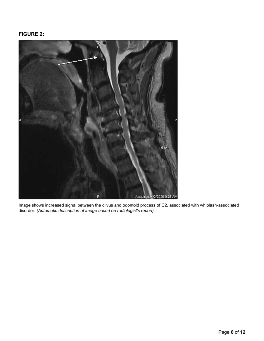
Drag the spread or use arrow keys to turn pagesSwipe the report or use the arrows to move one page at a time
Protected, de-identified preview. Your details go straight to our clinical team.
A fast mobile preview of the public report pages. The full protected walkthrough is still available from the sample report request flow.


Nine sections built for the people who actually read, use, and act on an imaging study. Every one designed to be the reason they come back.
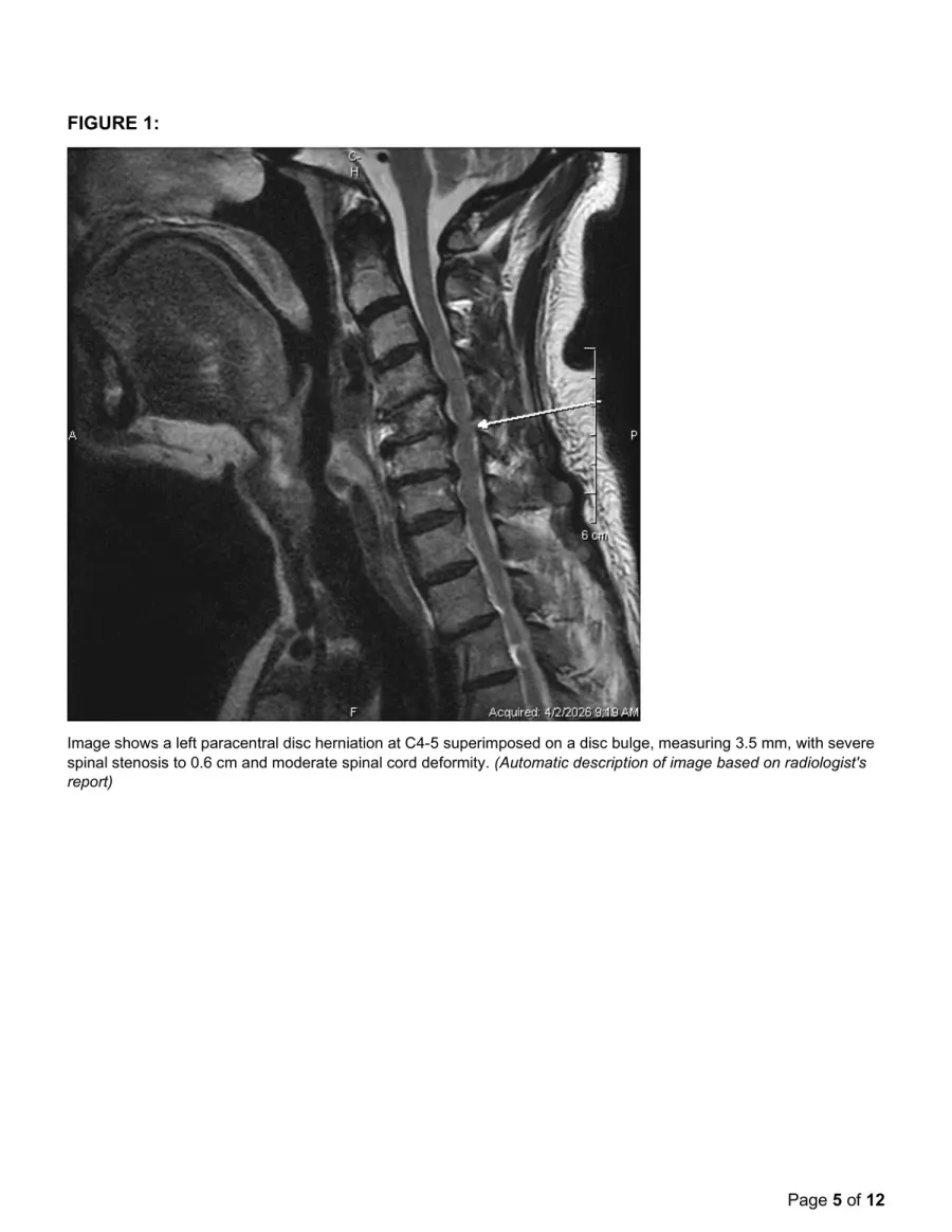
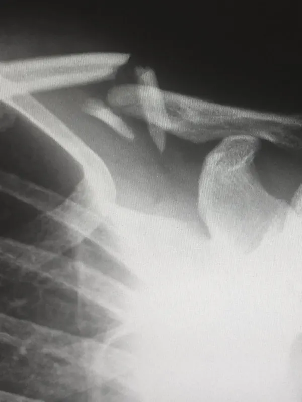
The actual MRI slice, with arrows pointing to the specific pathology. Findings stop being abstract. Patients, adjustors, and juries see the injury.
No extrapolation. No generic stock image. The annotation sits directly on the scan that was read.
Plain-language description below the figure, so any reader — clinical or lay — knows exactly what the arrow points to.
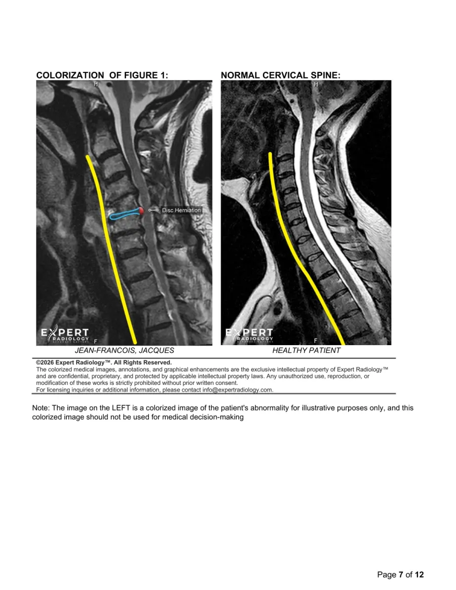
The patient's MRI beside a healthy patient of the same age. Spinal cord traced in yellow. Pathology circled in blue. The case tells itself.
The disc herniation is circled in blue. The spinal cord is traced in yellow. No squinting. No interpretation. Just evidence.
Instant clinical context. The jury does not need to know what a normal spine looks like. We show them.
Each report identifies the signing board-certified radiologist and relevant credentials. A real person. A real CV. A real deposition history.
Not a generalist worklist. Dr. Hewett signed this one. His credentials and fellowship are listed below his name.
Scan the code. See their training, certifications, and publications. Every radiologist vetted to the same standard.
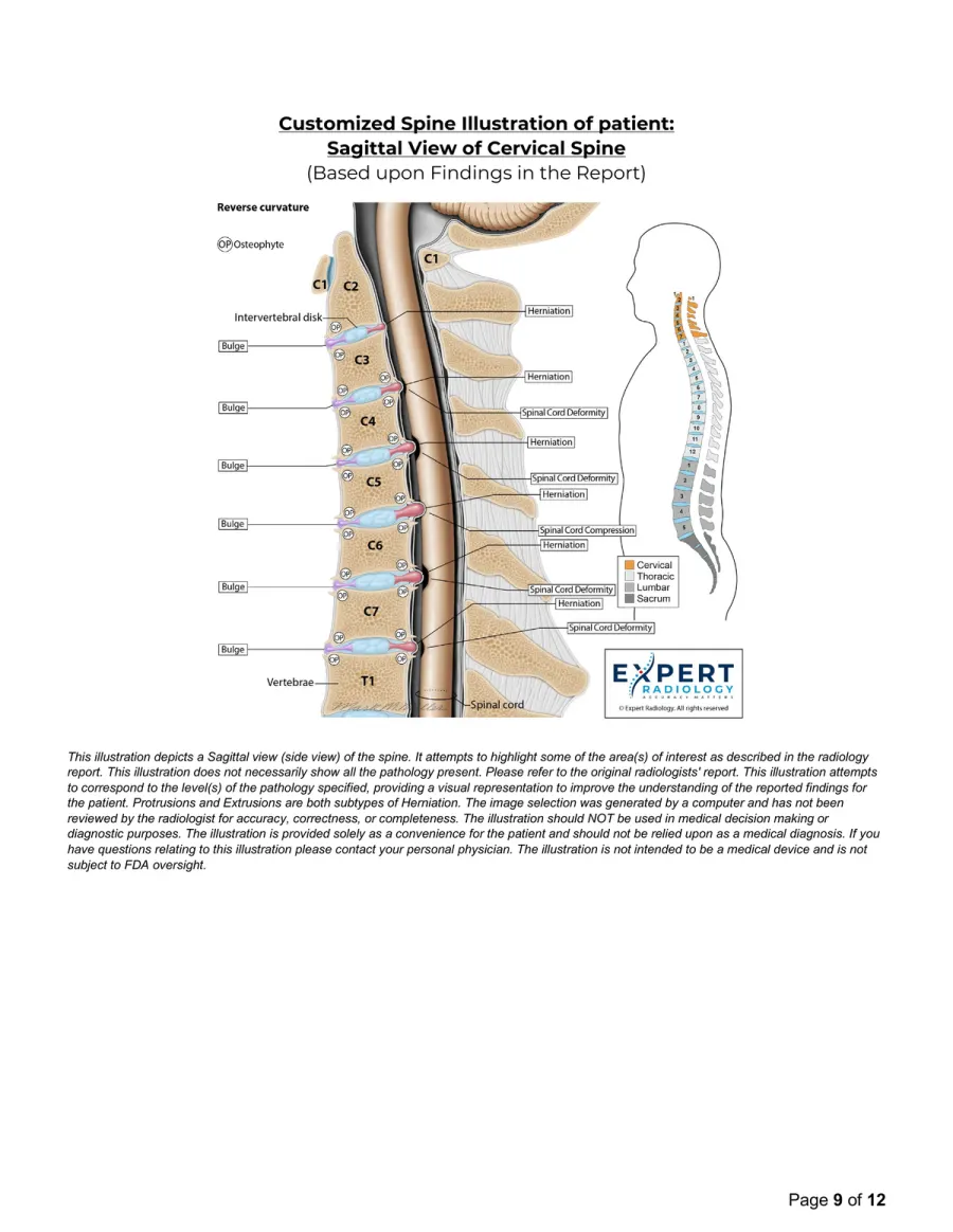
A custom illustration of the patient's spine, sagittal view, with every finding labeled at the exact vertebral level. The page a law firm can print poster-size for mediation.
C1 through T1, with findings called out in the margins. Bulges, herniations, spinal cord deformities, foraminal narrowing — nothing abstract.
The illustration reflects this patient's specific pathology at the specific level. A normal level looks normal. A herniation looks like a herniation.
A small full-body silhouette in the corner shows which region is illustrated. Context without clutter.
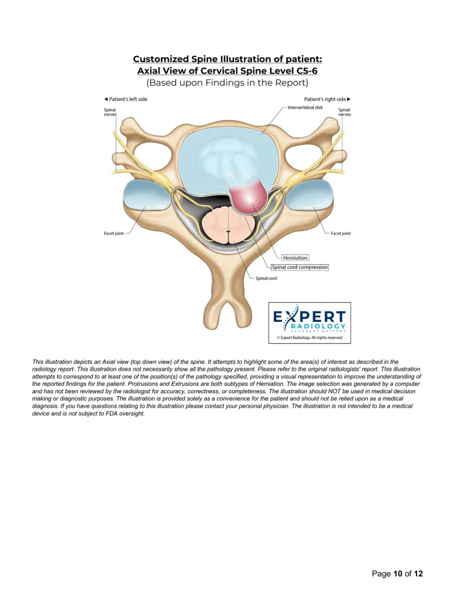
A top-down cross-section at the single most clinically significant level, showing exactly how the disc herniation impinges the spinal cord. Medical stories tell themselves.
For this case, C5-6. The disc herniation is colored pink. The cord compression is visible. Annotations label facet joints, spinal nerves, disc.
Every structure in the cross-section is labeled by name. No medical dictionary required to understand the injury.
Three core report moments, rendered as static mobile cards to avoid scroll-linked work on Chrome for iOS.
Arrows and callouts sit directly on the image being interpreted, so the finding is not abstract.
The pathology is circled and compared with normal anatomy so clinical and non-clinical readers see the difference.
Patient-specific anatomy pages show the injury at the exact level, ready for provider, patient, or legal review.
Book a 15-minute walkthrough. We'll show you how PrecisionPlus v3™ grows referral volume, cuts callback noise, and makes you the one providers, patients, and attorneys recommend by name.
We integrate with your PACS. Your studies route to the right subspecialist. Your referring providers get a report they bookmark.
Push studies to Expert Radiology directly from your PACS or submit through the ExRad Portal. No new workflow for your techs. Minimal disruption, go-live in days.
Routing is built around anatomy and modality. Neuro, MSK, spine, and body studies go to radiologists with relevant subspecialty experience.
Your referring providers get a PrecisionPlus v3™ report: colorized key images, custom illustrations, and a plain-language summary. Portal visibility keeps status clear from submission to delivery.
Whether you're an imaging center or provider, see how our subspecialty radiologists and PrecisionPlus v3™ reports serve your needs.
Same machines, different reports. Differentiate your center with illustrated, subspecialty-grade reports that eliminate callbacks and drive referral growth.

When patients see their injury illustrated, they can better understand the care conversation. Visual reports reduce confusion and support your plan of care.
See how imaging centers, providers, and legal teams use PrecisionPlus v3™ reports to make complex findings easier to understand, evaluate, and act on.
PrecisionPlus v3™ makes pathology unmistakable. Once the injury is clearly illustrated, text-only reports feel obsolete. Beyond the reports' clarity, their optimized workflow allows imaging centers to scale efficiently while maintaining the quality and credibility physicians and attorneys depend on.

These are the most effective MRI reports I've seen in my career. A true game changer for understanding and proving injury.

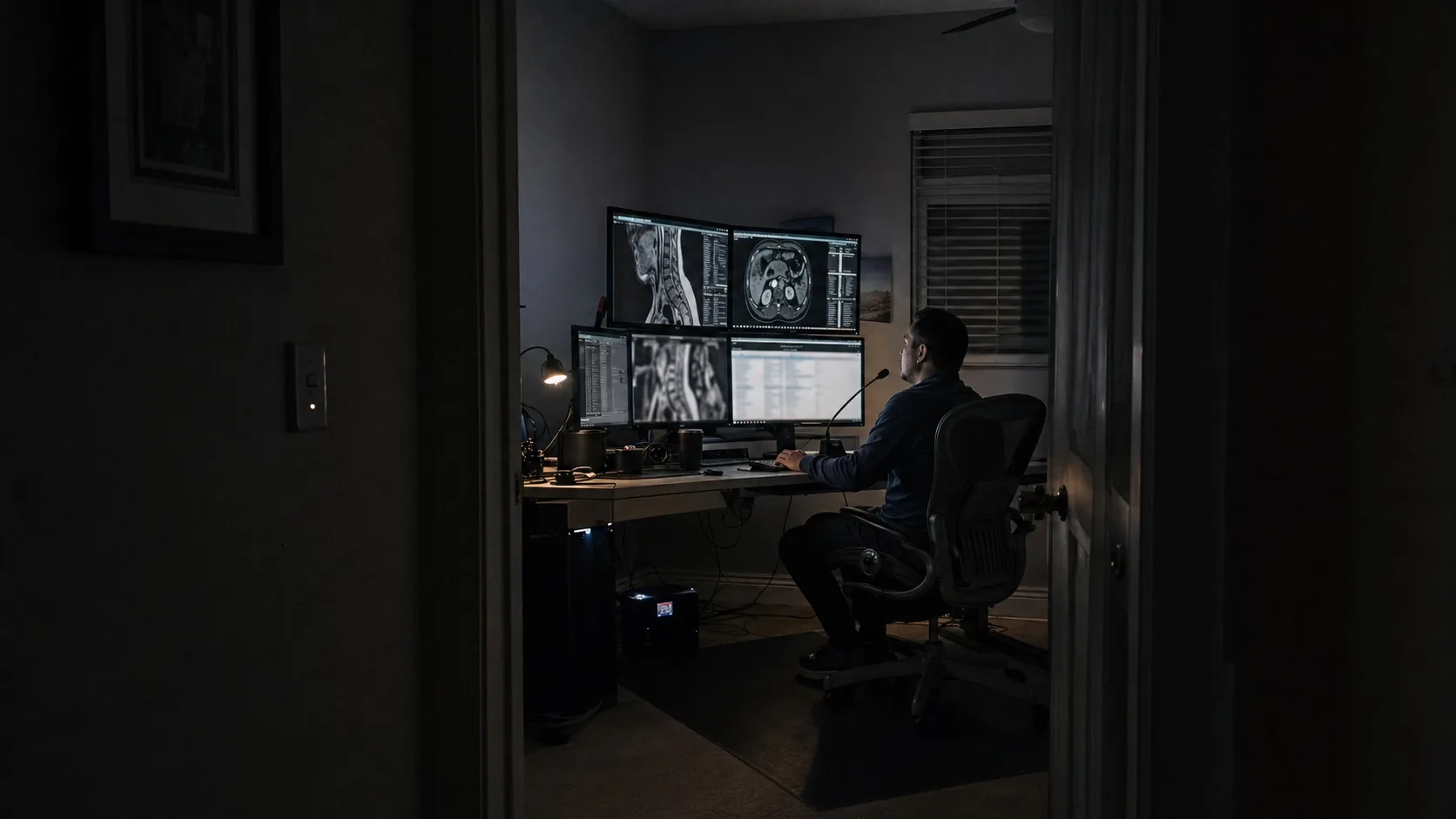
Coast Imaging has been working with the Expert Radiology team since February 2024. In January 2025, we upgraded to v3™ of their radiology reports, which added even more cutting-edge detail to the reports. After only a short few months, our volume has doubled, with very satisfied providers requesting more imaging than ever before.
The v3™ reports are incredibly helpful to transform raw images into clear, precise, and easy-to-understand pictures that have great evidentiary value. They are a great source of demonstrative evidence highlighting the findings, turning it into compelling testimony that is always grounded in science.

With Expert Radiology's support, we've been able to provide true concierge-level service to our patients and providers, leveraging their exceptional reports and optimized workflows to scale rapidly.

Our business has grown 27% in the four months since we started utilizing the v3™ report. We are quickly becoming the center of choice in our region.
Customer-reported individual result. Outcomes vary by market, operations, payer mix, competition, and referral strategy.
Everything you need to know about the report that changed radiology.
A standard report is text-only clinical jargon. PrecisionPlus v3™ adds colorized key images highlighting pathology, patient-specific custom medical illustrations, and a plain-language summary. The same subspecialty-grade read, delivered in a format that clinicians, attorneys, patients, and juries can immediately understand and act on.
Studies are routed to board-certified radiologists with relevant subspecialty expertise in neuroradiology, musculoskeletal imaging, body imaging, and related areas. Our team includes former academic faculty and physicians with decades of clinical and legal case experience.
PrecisionPlus v3™ is built for clear, decision-useful impressions. When imaging supports a specific finding, we describe it with measurements and relevant context. When imaging is genuinely ambiguous, the report says that directly instead of forcing certainty where the images do not support it.
Our reports are built with colorized key images, custom illustrations, and structured findings that legal teams commonly use in mediation, deposition preparation, and trial settings when admitted by the court. We also provide expert witness testimony and deposition support when the case fit and expert credentials align.
Turnaround depends on study complexity, clinical priority, current volume, and the agreed scope. Urgent work is prioritized appropriately, and case status remains visible through the ExRad Portal.
Our proprietary software generates pathology-specific medical illustrations tailored to each patient's findings. These are not generic anatomy diagrams. They are custom visuals that correspond directly to the pathology identified in the imaging study.
Every report includes: (1) detailed subspecialty interpretation with segmental analysis, (2) impressions with clear, clinically grounded confidence language, (3) annotated key MRI images, (4) colorized key images compared to normal anatomy, (5) custom medical illustrations in sagittal and axial views, (6) named reading radiologist signature with credentials, and (7) a simplified patient explanation. Reports are typically 8-11 pages.
Yes. Preview the protected report experience, or book a live demo and we'll walk you through a real report built for your specialty.
Book a 15-minute walkthrough. We'll show you how PrecisionPlus v3™ reports grow referral volume and make you the one providers, patients, and attorneys recommend by name.
A practical framework for imaging centers evaluating teleradiology providers, from turnaround time to subspecialty coverage and quality operations.