Generic stock

Matched anatomy, not matched findings. Useful as a reference, but not specific to the patient or the case.
Custom medical illustrations drawn from the patient’s actual scan, reviewed through the radiology workflow, and shipped inside every PrecisionPlus v3™ report. No clip-art. No outsourcing. No $900 exhibits three weeks late.


Other illustration workflows often start with clip art. Expert Radiology starts with the study, then builds the visual around the actual finding.
Matched anatomy, not matched findings. Useful as a reference, but not specific to the patient or the case.

Same anatomy, but built around the study findings and delivered as part of the report package.
Template anatomy diagram
The patient's actual study and report context
Generic labels that could fit any knee
Finding location, severity, and anatomy matched to the study
Visual reference only
Reviewed through the radiology workflow before delivery
Separate exhibit project
Included with the PrecisionPlus v3™ report package
General education
Report review, consults, mediation, and demand packages
Drag the center line left or right to compare a representative text-only format with PrecisionPlus v3™ visual reporting.
Drag the handle left or right to compare
Representative synthetic comparison for education only; not based on any specific competing radiology service.
Format
Language
Evidence
Turnaround
Browse by body region. Click any sample to open the same lightbox experience used on the free illustration page.
We can build shoulder visuals from the study and report context. Claim a free illustration and we will show you what your case looks like.
Request this anatomySend the case and we will confirm the right illustration approach for the finding.
Request this anatomyWant one made from your scan?
Claim Your Free IllustrationThe work starts with the study and ends inside the report, so the illustration supports the clinical read instead of floating beside it.
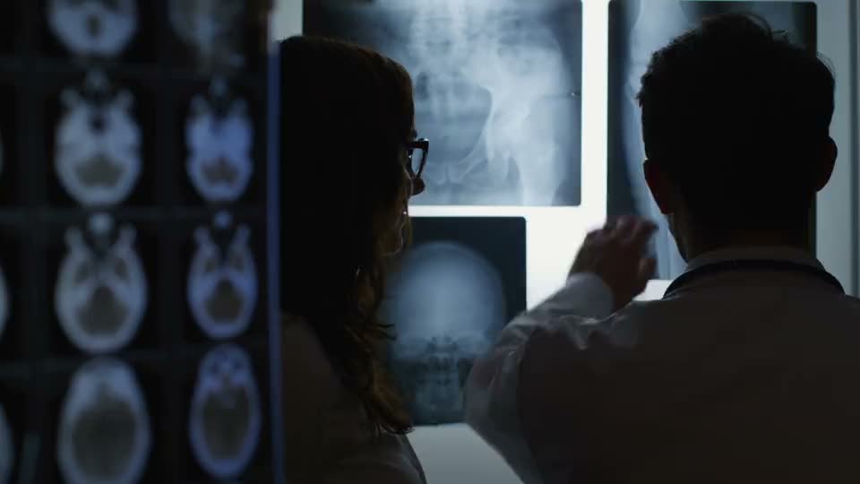
A subspecialty-focused reader reviews the study and flags the anatomy, level, and finding that need to be made clear.

The illustration is built to support the interpretation, so the image and written findings tell the same story.

Colorized key images and custom anatomy give the reader, provider, patient, or attorney a clearer way to review the finding.

The final asset is delivered with the PrecisionPlus v3™ report instead of handled as a separate exhibit project after the fact.

Whether you're an imaging center or provider, see how our subspecialty radiologists and PrecisionPlus v3™ reports serve your needs.
Same machines, different reports. Differentiate your center with illustrated, subspecialty-grade reports that eliminate callbacks and drive referral growth.

When patients see their injury illustrated, they can better understand the care conversation. Visual reports reduce confusion and support your plan of care.
One per practice. Case-specific and built from your actual scan. No credit card, no trial, no strings. Walk through four quick steps and you’ll have a legal-review-ready illustration from your own case.
Submit your details on the next page. Get a one-time ExRad Portal code instantly.
Your Imaging Workspace. Sign in with your code.
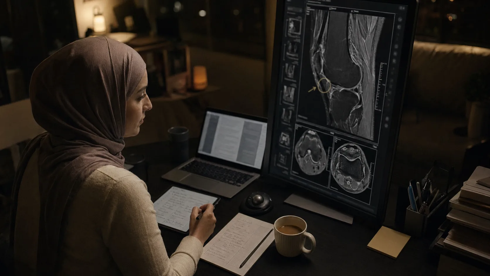
Drop in a de-identified DICOM, PDF report, or image file. HIPAA-compliant.
Built from your actual scan. Yours to use however you need.
HIPAA-compliant. De-identified uploads only. One per practice.
PrecisionPlus v3™'s illustrations were ready to use — we mounted them and walked the jury through the injuries. It saved time, reduced costs, and delivered real impact in the courtroom. If you're not using it for second opinions — or better yet, working with centers that offer it as a treating radiologist — you could be leaving significant value on the table.

When I try injury cases, I want the MRI to be PrecisionPlus v3™. The quality of the read and medical illustrations makes my client's injuries clear, credible, and impossible to ignore.

These are the most effective MRI reports I've seen in my career. A true game changer for understanding and proving injury.

PrecisionPlus v3™ makes pathology unmistakable. Once the injury is clearly illustrated, text-only reports feel obsolete. Beyond the reports' clarity, their optimized workflow allows imaging centers to scale efficiently while maintaining the quality and credibility physicians and attorneys depend on.

The v3™ reports are incredibly helpful to transform raw images into clear, precise, and easy-to-understand pictures that have great evidentiary value. They are a great source of demonstrative evidence highlighting the findings, turning it into compelling testimony that is always grounded in science.

Everything you need to know about the illustrations that ship with every PrecisionPlus v3™ report.
Spine, knee, shoulder, brain, and soft-tissue illustrations. Every view that matters to the case: sagittal, axial, frontal, and exploded anatomical views. Delivered as high-resolution PNG, print-ready PDF, or embedded directly inside the PrecisionPlus v3™ report.
Medical illustrators trained in anatomical precision produce every draft. Illustrations are reviewed through the radiology workflow for anatomical consistency and clinical relevance before delivery.
Always custom. Each illustration is built from the patient's actual imaging findings: the specific anatomy, level, mechanism, and severity. Stock anatomy diagrams do not meet the "fair representation" standard that courts and clinicians expect.
They can support case review, mediation preparation, deposition preparation, or trial exhibits when the appropriate expert and court permit their use. The visuals are built from the patient's actual imaging findings and reviewed through the radiology workflow. Dr. Knapp has participated in over 400 depositions tied to illustrated radiology reports.
Production tools may accelerate drafting and image preparation, but the final illustration is reviewed and verified by a human board-certified radiologist before it ships. Admissibility and exhibit use depend on jurisdiction, court rules, case facts, methodology, and the supporting expert.
Timing depends on case complexity and current report volume. Because illustrations ship inside the report rather than as standalone legal exhibits, they follow the report workflow instead of the traditional 3 to 4 week medical-legal illustration process.
No. Every PrecisionPlus v3™ report includes custom illustrations. Report Enhancement adds them to outside reports. For reference, the industry average is roughly $900 per illustration in personal injury work.
HIPAA-compliant infrastructure, de-identified uploads, encrypted storage. The ExRad Portal workflow follows the same security posture as the rest of our stack.
Book a 15-minute walkthrough. We’ll show you how illustrations inside PrecisionPlus v3™ grow referral volume and make you the one providers, patients, and attorneys recommend by name.
A second read can change everything. Explore how radiology second opinions catch missed findings and strengthen clinical and legal outcomes.