
In the consult
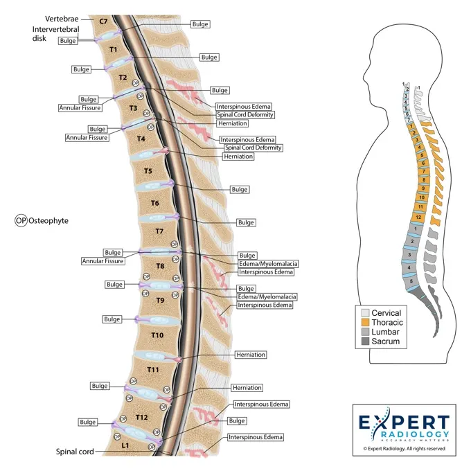
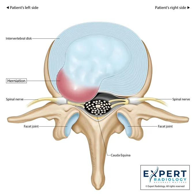
Colorized spinal anatomy
Point to the exact disc, the exact nerve root, the exact facet. Patients see what your adjustment is targeting and understand the whole plan.
"I can actually see what's wrong."What patients say
Patients can nod through an MRI explanation and still leave without a clear mental picture of what a disc bulge actually looks like. Visual reports close the gap between your care plan and their understanding with an image they can revisit later.
A care plan you can't see is a care plan patients stop believing in. By the third visit, urgency fades.
A colorized v3™ report reinforces your plan every time the patient looks at it between visits.
"L5-S1 disc desiccation with broad-based bulge" does not explain the anatomy in a way most patients can revisit later.
The disc compression, foraminal narrowing, and facet degeneration each point to a specific part of your care conversation.
Patients shop you against the next office. You need a reason they pick you and stay.
v3™ becomes a reason referring attorneys, PCPs, and past patients send work to your practice.
Every PrecisionPlus v3™ report is built so your patients can see the relevant imaging findings and understand why your care plan matters.
Subspecialty-focused spine and MSK reads paired with custom medical illustrations, colorized key images, and a plain-language patient summary. One document with portal tracking, ready to walk a chiropractic patient through in the consult.
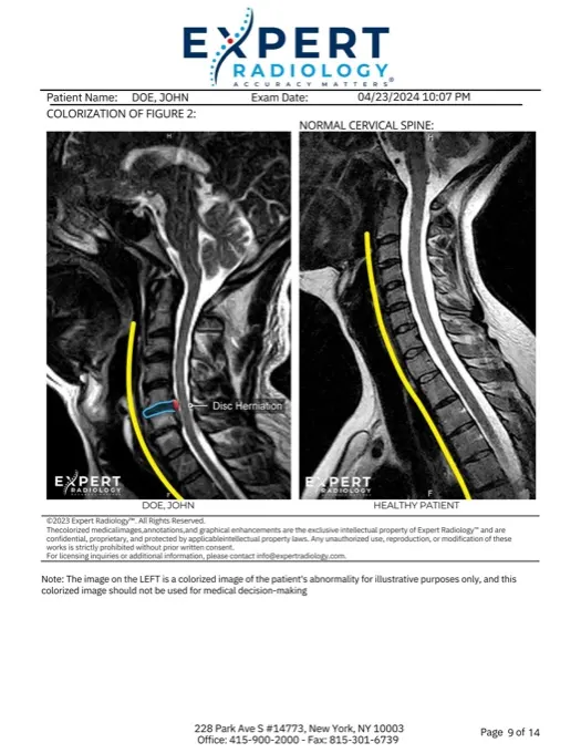
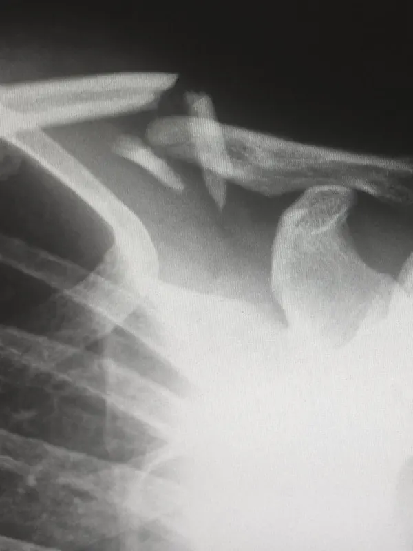
Findings highlighted directly on the imaging with color-coded annotations. Point to the screen during the consult, and patients see the pathology immediately.
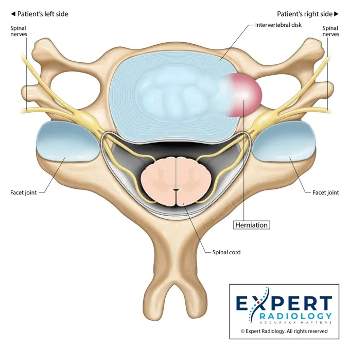
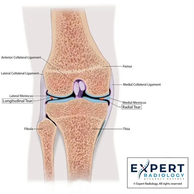
Anatomical illustrations created for each case. One illustration does more than two pages of text, and patients understand their condition in seconds.
A summary patients can actually read, understand, and take home. It reinforces your care-plan explanation between visits, when they need it most.
Studies route by anatomy, modality, and available subspecialty expertise across spine, MSK, neuro, and body imaging.
Clear impressions with appropriate confidence language. When the imaging supports a finding, we describe it directly; when it is genuinely ambiguous, we say that too.
Status stays visible from submission to delivery, so your team knows where the report stands before the next patient conversation.
Visual reports reinforce the care plan at the table and after the patient leaves, so the reason for care stays clear.
Point to the exact disc, the exact nerve root, the exact facet. Patients see what your adjustment is targeting and understand the whole plan.
"I can actually see what's wrong."What patients say
A take-home patient summary that explains why weeks of adjustments, decompression, or rehab matter. They read it between visits and come back ready.
"My wife read it too."What patients say
Subspecialty-focused reads built to surface disc protrusions, foraminal stenosis, and facet arthropathy with the detail your care plan needs.
"This read is so much more thorough."What patients say
Three steps, minimal workflow disruption. Use your existing imaging center or one from our network.
Send the existing study through the ExRad Portal or your imaging center's workflow. No new software, no new logins.
Subspecialty-focused read, colorized key images, and a plain-language patient summary, with status visible through delivery.
Walk the patient through the report in the consult and send them home with visual context they can revisit.
Everything you need to know about v3™ reports and patient education.
We hand you a document that the patient can follow along with: colorized key images, anatomical callouts, and a plain-language summary. You stop explaining medical terminology and start walking them through their own imaging.
Yes. The same visual documentation that supports patient education can also make demand letters and mediation exhibits easier to evaluate.
v3™ is additive. We enhance the read you already get, or we do the primary subspecialty read ourselves. Either way your patients leave with something no other chiropractic office is handing out.
Turnaround depends on case complexity, clinical priority, current volume, and the agreed workflow. Urgent cases are prioritized appropriately.
Request a sample v3™ report and see the document your patients take home instead of a wall of grayscale and medical jargon.
Estimate how much faster your reports could arrive with Expert Radiology. Enter your current volume and see projected turnaround improvements.