
The language of imaging,
in plain terms.
Clear definitions of the radiology, MRI, teleradiology, and personal-injury terms that show up in reports and case files. Written so anyone, not just a radiologist, can follow them. If a term you need is missing, contact us and we will add it.
Imaging and technology
- Magnetic Resonance Imaging (MRI)
-
MRI is a scan that uses a strong magnetic field and radio waves, not radiation, to produce detailed images of soft tissue such as the brain, spine, joints, and ligaments. It is the modality of choice for many injuries because it shows soft-tissue damage that X-ray and CT can miss.
- Teleradiology
-
Teleradiology is the practice of transmitting medical images to a radiologist in another location for interpretation. It lets an imaging center reach subspecialty reads without employing every specialty in-house, with studies routed through the center’s existing workflow and reports returned to its system.
How teleradiology works - PACS
-
A Picture Archiving and Communication System (PACS) is the software an imaging facility uses to store, retrieve, and share medical images. Teleradiology partners connect to a center’s PACS so studies route out for reading and finished reports return without a separate workflow.
For imaging centers - RIS
-
A Radiology Information System (RIS) is the software that manages a radiology operation’s scheduling, patient tracking, billing, and reporting. It works alongside the PACS, which stores the images, to run the operational side of imaging.
- DICOM
-
DICOM (Digital Imaging and Communications in Medicine) is the standard format and protocol for storing and exchanging medical images. Because scanners, PACS, and viewers all speak DICOM, an MRI acquired on one system can be read on another, which is what makes teleradiology and second opinions possible.
- Modality
-
In imaging, a modality is a type of scan or the equipment that produces it: MRI, CT, X-ray, ultrasound, and others. Each modality shows different tissue best, so the right one depends on the clinical question and the suspected injury.
- Subspecialty radiology
-
Subspecialty radiology is the practice of a radiologist focusing on one area of the body or type of imaging, such as neuroradiology, musculoskeletal, or body imaging. Concentrated volume in one anatomy builds pattern recognition that a generalist reading everything cannot match.
What is subspecialty radiology - Subspecialist
-
A subspecialist is a radiologist who has trained and concentrated in a specific anatomy or imaging type. Matching a study to a subspecialist in that exact area, rather than a generalist, is how subtle or complex findings are most reliably caught and described.
Meet our radiologists
Reports and reading
- PrecisionPlus v3™ report
-
A PrecisionPlus v3™ report is Expert Radiology’s illustrated MRI report. It pairs a subspecialty radiologist’s findings with colorized key images and patent-pending custom illustrations, plus a plain-language explanation, so patients, providers, and legal teams can understand the injury without a medical background.
See the PrecisionPlus v3™ report - Colorized key images
-
Colorized key images are the patient’s own scan images with the relevant finding highlighted in color and contrasted with normal anatomy. They turn a grayscale image that only a radiologist can read into something a layperson can see and understand at a glance.
Inside a v3 report - Custom medical illustration
-
A custom medical illustration is a drawing built from the patient’s actual imaging that shows the specific injury on the relevant anatomy. Unlike stock art, it reflects this patient’s findings, which makes complex pathology clear to laypeople reviewing the case.
Custom medical illustrations - Report enhancement
-
Report enhancement, sometimes called a V3 overlay, adds colorized images, custom illustrations, and a plain-language explanation to an existing radiology report from another radiologist. It upgrades the readability of a report without re-reading the study from scratch.
Report enhancement - Second opinion
-
A radiology second opinion is an independent re-read of an existing study by another radiologist, ideally a subspecialist in that anatomy. The reviewer works from the original images rather than the original report and issues new findings, which can confirm, refine, or contradict the first read.
Request a second opinion - Turnaround time (TAT)
-
Turnaround time is the interval between when a study is submitted and when the finalized report is returned. It depends on study type, volume, and clinical priority. Expert Radiology scopes turnaround at intake, keeps case status visible, and prioritizes urgent reads.
More in our FAQ - Hedge words
-
Hedge words are non-committal phrases in a radiology report such as cannot exclude, clinical correlation recommended, or may represent. They manage uncertainty between physicians, but to a non-medical reader they can read as doubt and weaken how clearly a finding is understood.
- Credentialing
-
Credentialing is the process of verifying a radiologist’s licenses, board certification, training, and history before they read studies for a facility or in a given state. Maintaining current credentialing files is what allows reads to be performed compliantly across many states.
Legal and case work
- Demand package
-
A demand package is the documented account a legal team assembles to present a claim, combining the liability narrative, medical records, imaging, billing, and the impact on the person. It is built to persuade a non-medical reader, which is why clear imaging evidence matters so much.
Anatomy of a Demand Package - Defensible report
-
A defensible report is one whose findings are authored and signed by a credentialed radiologist, tied to the specific images, and stated using standard criteria, so they hold up under scrutiny. Visual elements illustrate documented findings rather than adding claims the images do not support.
For legal teams - Expert witness
-
In radiology, an expert witness is a credentialed radiologist who can explain imaging findings in a legal setting. Whether and how that testimony is used depends on the case, the retained expert, and the court. The imaging report itself is documentation, not testimony.
Expert witness services - Medical lien
-
A medical lien is an arrangement in which a provider treats a patient on a personal-injury claim and is paid from the eventual settlement rather than up front. Lien-based care depends on clear documentation of injury, which makes legible imaging evidence valuable.
For medical lien partners - Causation
-
In injury cases, causation is the link between the incident and the documented injury. Imaging supports causation when findings, acuity markers, and comparison with prior studies help distinguish a new injury from pre-existing or incidental changes. It is a clinical and legal judgment, not a single image.
How we support legal teams
Common MRI findings
- Disc herniation
-
A disc herniation occurs when the soft center of a spinal disc pushes through its outer wall, sometimes pressing on nearby nerves. On MRI it is described by level, for example L4-L5, and by type, and it is one of the most common findings in spine injury cases.
- Spinal stenosis
-
Spinal stenosis is a narrowing of the spinal canal or the openings where nerves exit, which can compress the spinal cord or nerve roots. MRI shows where and how severely the space is narrowed, which helps connect imaging to a patient’s symptoms.
- Radiculopathy
-
Radiculopathy is the set of symptoms, such as pain, numbness, or weakness, that occurs when a spinal nerve root is compressed or irritated, often by a disc herniation or stenosis. Imaging identifies the structural cause that may explain the reported symptoms.
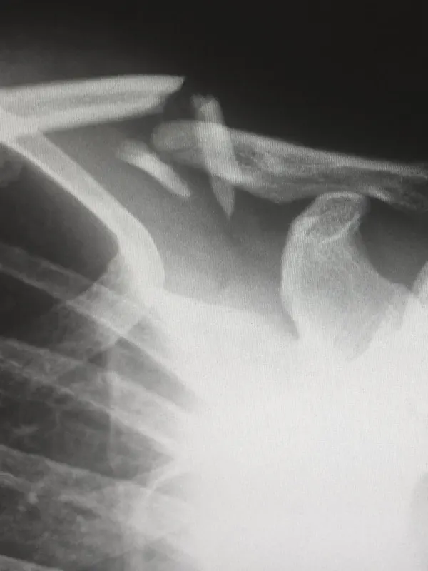
- Rotator cuff tear
-
A rotator cuff tear is a tear in one of the tendons that stabilize the shoulder. MRI distinguishes a partial tear from a full-thickness tear and shows its size and location, detail that matters for treatment decisions and for documenting a shoulder injury.
See the terms in a real report.
The fastest way to understand an illustrated MRI report is to see one. Book a walkthrough or send us a study for a second opinion.