
Understanding MRI vs CT for Injury Cases
When is an MRI the right call versus a CT scan? We break down the clinical scenarios, strengths, and limitations of each modality for injury and trauma imaging.

Table of Contents
- The Fundamental Difference: What Each Modality Actually Sees
- Spine Injuries: Where MRI Dominates
- Traumatic Brain Injury: The CT Blind Spot
- Orthopedic Injuries: Fractures, Ligaments, and the Occult
- The Cost Reality: MRI vs CT Pricing
- Impact on Personal Injury Case Value
- Who Reads It Matters More Than What You Order
- When CT Wins: The Right Tool for the Right Question
- The Dual-Modality Approach: Best Practice for Injury Cases
- The Bottom Line
Key Takeaways
- MRI detects disc herniations with 91.7% sensitivity versus 55-83.3% for CT, making it the definitive soft tissue modality for spine injury cases.
- CT scans miss 50-80% of diffuse axonal injuries in traumatic brain injury. If your client had a head impact, an MRI is not optional.
- Subspecialty radiologist error rates run 2.0-2.7%, compared to 12.4-40% for generalists. The reader matters as much as the modality.
- Public settlement/verdict benchmarks vary widely and should not be used as guarantees; imaging documentation is best evaluated case by case.
- The cost gap between MRI (~$1,325) and CT (~$525) is real, but choosing the wrong modality can create missed diagnoses, delays, and weaker documentation.
Every week, someone asks the same question: "Should I order an MRI or a CT?" The honest answer is that it depends on what you are looking for. But in injury cases where soft tissue damage and early documentation matter, the answer skews heavily toward MRI. Not always. But more often than most people realize.
This article lays out the evidence. Not marketing claims. Not opinions dressed as facts. Actual sensitivity data, cost comparisons, and outcome research so you can make informed decisions about imaging for injury cases.
The Fundamental Difference: What Each Modality Actually Sees
CT and MRI are not interchangeable. They use completely different physics to produce images, and those differences determine what each one reveals.
CT (computed tomography) uses X-ray beams rotated around the body to generate cross-sectional images. It excels at dense structures: bone, calcifications, acute bleeding. A CT scan takes seconds. That speed saves lives in the emergency department when you need to rule out a skull fracture or internal hemorrhage at 2 AM.
MRI (magnetic resonance imaging) uses powerful magnetic fields and radiofrequency pulses to map hydrogen atoms in tissue. Because different tissues contain different amounts of water, MRI produces extraordinary contrast between soft tissue structures. Ligaments, tendons, cartilage, intervertebral discs, the spinal cord, brain parenchyma: MRI shows them all in detail that CT simply cannot match.
Think of it this way. CT is a flashlight in a dark room. It illuminates hard edges and sharp angles brilliantly. MRI is a thermal camera. It reveals the things hiding in the shadows: the swelling, the torn fibers, the subtle signal changes that tell you tissue has been damaged even when the bones look fine.
Spine Injuries: Where MRI Dominates
The spine is where the MRI vs. CT debate is most lopsided. A 2022 meta-analysis published in Frontiers in Surgery compared the two modalities for lumbar disc herniation detection. The results were not close. MRI achieved 91.7% sensitivity. CT ranged from 55% to 83.3%.1
That gap matters in real cases. A disc herniation missed on CT might mean a client is labeled as having only a "soft tissue strain." With MRI and a subspecialty read, the record may document pathology that was not visible on CT. The imaging modality alone does not create the injury; it can reveal it.
The ACR Appropriateness Criteria (2024 edition) reinforces this. For suspected spinal cord injury, ligamentous injury, and disc pathology, MRI is rated "usually appropriate" while CT is recommended primarily for acute bony evaluation.4 The radiology community is not ambiguous about this.
Traumatic Brain Injury: The CT Blind Spot
CT is the first-line imaging for acute head trauma. It is fast, widely available, and excellent at detecting skull fractures and acute intracranial hemorrhage. Nobody disputes that.
The problem is what CT misses. A 2024 systematic review comparing MRI and CT for traumatic brain injury (TBI) found MRI sensitivity of 0.91 versus 0.82 for CT.2 That 9-point gap represents real patients with real injuries that CT did not detect.
The gap widens dramatically for diffuse axonal injury (DAI), one of the most serious consequences of traumatic brain injury. CT misses 50-80% of diffuse axonal injuries.8 Fifty to eighty percent. These are patients with documented loss of consciousness, cognitive decline, and persistent neurological symptoms whose CT scans look normal. Without a follow-up MRI, their injuries are invisible to the medical record, to insurance adjusters, and to juries.
For attorneys handling motor vehicle accident cases with head impact: if the ER CT was "negative" but your client is still struggling with headaches, memory problems, or personality changes six weeks later, that negative CT does not rule out brain injury. It rules out hemorrhage and fracture. An MRI with diffusion-weighted imaging (DWI) and susceptibility-weighted imaging (SWI) sequences is the next step. Often, it changes everything.
Orthopedic Injuries: Fractures, Ligaments, and the Occult
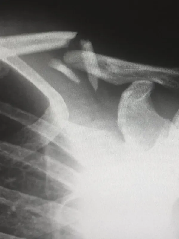
Here is where the picture gets nuanced. CT is excellent for fractures. Full stop. If you suspect a complex fracture, CT will map every fragment with sub-millimeter precision. For surgical planning, CT is often essential.
But not every fracture is visible on CT. For stress fractures, MRI sensitivity ranges from 68-99% compared to just 32-38% for CT.7 The reason: MRI detects the bone marrow edema (swelling within the bone) that precedes a visible fracture line. CT only sees the fracture once it is structurally complete.
Occult hip fractures tell a similar story. A 2024 meta-analysis found MRI to be superior for detecting fractures that initial imaging missed entirely. In the study data, CT missed 12 hip fractures that ultimately required surgery.3 Twelve patients who needed operations that CT said were not necessary. That is not a rounding error.
For ligament injuries, there is no contest. The ACL is the most-studied example: MRI achieves 87% sensitivity and 93% specificity for ACL tears.7b CT cannot reliably visualize ligaments at all. The same applies to meniscal tears, rotator cuff injuries, and virtually every other ligamentous or tendinous pathology relevant to personal injury cases.
The Cost Reality: MRI vs CT Pricing
Cost is a legitimate consideration. Let's be straightforward about the numbers.
| Factor | MRI | CT |
|---|---|---|
| Average cost | ~$1,325 | ~$525 |
| Price range | $400 - $12,000 | $300 - $6,750 |
| Hospital vs. independent | Hospital-based facilities charge 2-5x more than independent centers | |
| Scan time | 20-60 minutes | 5-15 seconds |
| Radiation exposure | None | Yes (ionizing radiation) |
Source: GoodRx Health, 2024 pricing data.10
Yes, MRI costs roughly 2.5x more than CT on average. But context matters. The question is not "which scan is cheaper?" The question is "what will this scan reveal, and what is the cost of missing something?"
A $525 CT that misses a disc herniation or ligament tear is not a savings. It is a delay. The patient still has the injury. The attorney still needs the evidence. You end up ordering the MRI anyway, weeks or months later, after the acute inflammatory changes have started resolving and the causation timeline has weakened.
Impact on Injury Documentation
This is where imaging selection stops being only a clinical question and becomes a documentation question. The data needs careful framing.
Some legal-market sources report large differences in settlement and verdict outcomes when structural pathology is documented by MRI, but those figures are not clinical benchmarks and should not be treated as outcome guarantees.9 The safer takeaway is simpler: objective MRI findings can materially change the documentation available for case review.
Public verdict summaries for disc-herniation cases vary widely by jurisdiction, injury severity, causation record, and case facts.9 Imaging does not determine an outcome by itself, but a clearer MRI record gives reviewers more objective information to evaluate.
"MRI within 72 hours of the incident creates the strongest causation baseline. It captures acute inflammatory changes, edema, and hemorrhage that fade with time. Every week of delay gives the other side another argument."
Timing matters as much as modality. Early MRI may create a clearer contemporaneous baseline when acute bone marrow edema, ligamentous edema, or hemorrhagic components are visible. Wait three months, and some acute findings may resolve. Now the discussion may shift toward chronic changes, timing, and pre-existing-condition arguments.
Want to model the documentation value for your workflow? Our Legal Imaging Value Calculator lets you plug in your matter volume, review time, exhibit-prep, and report-cost assumptions.
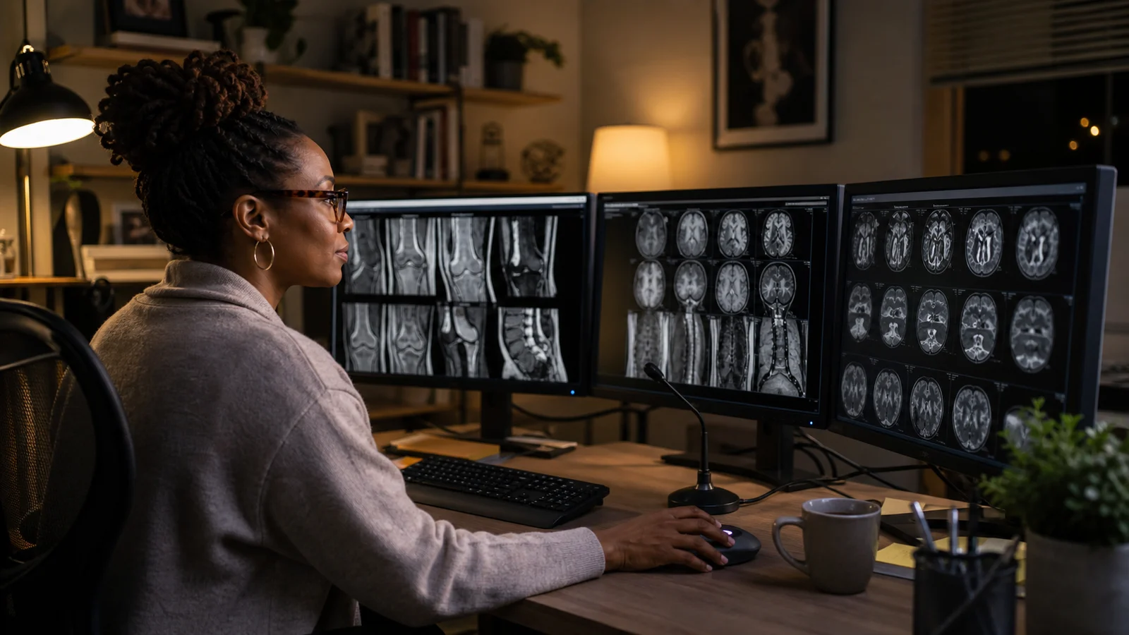
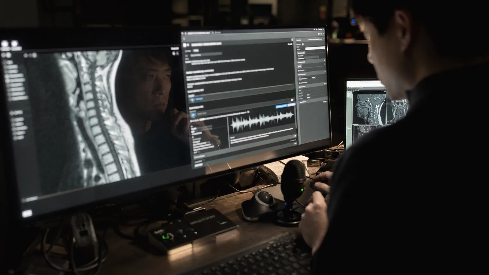
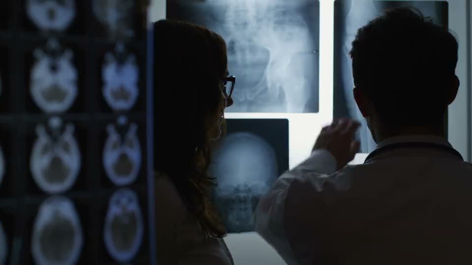
Who Reads It Matters More Than What You Order
Here is something most people outside radiology do not appreciate: ordering the right scan is only half the equation. The other half is who interprets it.
A 2017 study by Brady published in the British Journal of Radiology quantified radiologist error rates. General radiologists demonstrated error rates between 12.4% and 40%. Fellowship-trained subspecialists: 2.0-2.7%.6 Read those numbers again. The gap between a generalist and a subspecialist can be 15x in diagnostic accuracy.
A landmark 10-center spine MRI study by Herzog and colleagues, published in The Spine Journal (2017), sent the same patient to 10 different imaging centers. The result: zero unanimous findings across the 10 reads, and a 43.6% miss rate for significant pathology.5 Same patient. Same spine. Same pathology. Ten different answers.
This is why we emphasize subspecialty radiology so aggressively. An MRI read by a generalist who misses the finding is worse than not ordering the MRI at all, because now you have a "negative" scan in the record that the other side will use against your client.
The solution is straightforward: order the right study and make sure it is read by the right specialist. If the initial read looks incomplete or uses ambiguous language like "cannot exclude" or "correlation recommended," a subspecialty second opinion can clarify the record. The practical value depends on the matter and the team using the documentation.
When CT Wins: The Right Tool for the Right Question
This article is not anti-CT. CT is indispensable in the right clinical context. Dismissing it would be intellectually dishonest. Here is when CT is the correct first choice:
- Acute trauma evaluation. Patient arrives in the ER with multi-system injuries. CT of the head, chest, abdomen, and pelvis in under 60 seconds. MRI takes 20-60 minutes per body part. In an emergency, speed wins.
- Complex fracture mapping. Comminuted fractures, facial bone fractures, pelvic ring injuries: CT with 3D reconstruction gives surgeons the roadmap they need for operative planning.
- Acute intracranial hemorrhage. In the first 24 hours after head trauma, CT is faster and highly sensitive for bleeding that requires emergency intervention.
- Patients who cannot undergo MRI. Certain cardiac pacemakers, metallic implants, severe claustrophobia, or body habitus that exceeds scanner limits. CT remains the fallback.
- Lung and chest pathology. CT angiography for pulmonary embolism, high-resolution CT for lung parenchyma. MRI has limited utility in the chest.
The key principle: CT answers "is anything broken or bleeding right now?" MRI answers "what is the full extent of the injury?" Both questions matter. They just matter at different times.
The Dual-Modality Approach: Best Practice for Injury Cases
In most personal injury cases, the optimal strategy uses both modalities sequentially:
| Phase | Modality | Purpose | Timing |
|---|---|---|---|
| 1 | CT | Rule out fractures, hemorrhage, acute emergencies | Day of injury (ER) |
| 2 | MRI | Full soft tissue evaluation, establish causation baseline | Within 72 hours |
| 3 | Subspecialty read | Precise characterization of findings, legal-review-ready report | As soon as images are available |
This sequence gives you the best of both worlds. The CT handles the emergency triage. The MRI builds the diagnostic foundation. And the subspecialty interpretation ensures nothing is missed, minimized, or lost in vague language.
For attorneys handling injury cases, this approach can produce a stronger evidentiary record. You have the acute CT showing emergency triage, the early MRI capturing tissue-level damage while it is still fresh, and a subspecialist report that characterizes findings in clear language with appropriate confidence.
The Bottom Line
CT and MRI are complementary, not competing. But for soft tissue injuries, brain injury beyond acute hemorrhage, ligament tears, disc pathology, and occult fractures, MRI is the superior modality. The sensitivity data is consistent across decades of research.
The cost difference between modalities is real but often misleading. The true cost is not only what appears on the bill. It can also be the missed diagnosis, delayed care conversation, or undocumented injury. Whether an MRI is worth the expense depends on the clinical question and the documentation need.
And regardless of which modality you choose, the interpreter is the variable that determines whether the study is useful or wasted. A 43.6% miss rate across 10 imaging centers is not a failure of MRI technology.5 It is a failure of interpretation. Subspecialty reads close that gap.
Order the right study. Get it read by the right specialist. Do it early. That three-part formula sounds simple because it is. The hard part is finding a radiology partner who delivers on all three.
Sources
- Lumbar disc herniation imaging meta-analysis. Frontiers in Surgery, 2022. MRI sensitivity 91.7% vs CT 55-83.3% for disc herniation detection. frontiersin.org
- Comparative efficacy of MRI and CT for traumatic brain injury: a systematic review. PMC, 2024. MRI sensitivity 0.91 vs CT 0.82. ncbi.nlm.nih.gov
- Occult hip fracture meta-analysis: MRI vs CT comparative performance. PMC, 2024. CT missed 12 fractures requiring surgical intervention. ncbi.nlm.nih.gov
- ACR Appropriateness Criteria: Suspected Spine Trauma, 2024 edition. American College of Radiology. acsearch.acr.org
- Herzog R, Elgort DR, Flanders AE, Moley PJ. Variability in diagnostic error rates of 10 MRI centers performing lumbar spine MRI exams on the same patient within a 3-week period. The Spine Journal, 2017;17(4):554-561. pubmed.ncbi.nlm.nih.gov
- Brady AP. Error and discrepancy in radiology: inevitable or avoidable? Insights into Imaging (British Journal of Radiology), 2017;8(1):171-182. General error rate 12.4-40%; subspecialist 2.0-2.7%. pubmed.ncbi.nlm.nih.gov
- MRI vs CT for stress fracture detection. PubMed, 2015. MRI sensitivity 68-99% vs CT 32-38%.
- ACL tear diagnostic accuracy meta-analysis. PubMed, 2016. MRI sensitivity 87%, specificity 93%. pubmed.ncbi.nlm.nih.gov
- Diffuse Axonal Injury. StatPearls, National Library of Medicine (NIH). CT misses 50-80% of DAI. ncbi.nlm.nih.gov
- Edwards Patterson Law. Public injury-case settlement and verdict discussion involving MRI documentation. Legal-market benchmarks vary widely and should not be treated as guarantees. edwardspattersonlaw.com
- GoodRx Health. MRI and CT scan cost comparison data, 2024. Average MRI ~$1,325 ($400-$12,000); average CT ~$525 ($300-$6,750). goodrx.com