


Which tract. Not just that something’s off.
Advanced DTI measures up to 33 major white-matter tracts, one at a time, against a normative range matched for age and sex. Any tract outside its range is flagged, and a board-certified neuroradiologist reads the study and signs the report.
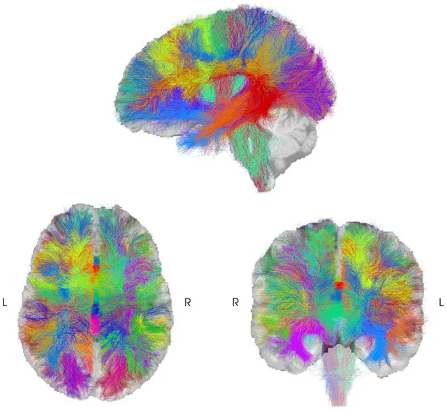
- Up to 33
- major white-matter tracts, each measured on its own
- Age and sex matched
- normative comparison for every tract
- FDA 510(k) cleared
- IMEKA’s ANDI analysis software
A whole-brain DTI read can tell you connectivity looks abnormal. Advanced DTI tells you where.
-
Up to 33 tracts, measured one by one.
Each major white-matter tract gets its own result, so a finding has a name and a place.
-
Compared to the right reference.
Every tract is checked against a normative range matched for the patient’s age and sex.
-
Flags you can find.
Any tract outside its range is marked in the report, with the measurement behind the flag.
-
The full IMEKA/ANDI report.
The complete tractography report is included with every Advanced DTI study.
-
A summary patients can read.
A computer-generated, plain-language summary explains the tractography findings for the patient. It is not reviewed by the radiologist, and it never replaces the radiologist’s report.
-
Read and signed by a neuroradiologist.
A board-certified neuroradiologist reviews the analysis alongside the conventional MRI and signs the report.
Two levels of analysis, in every Advanced DTI report.
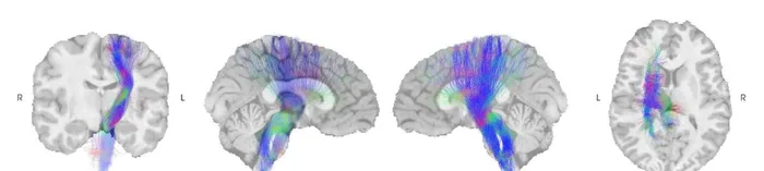
Tract-level detail
Each measured bundle is rendered on the study it came from, so the finding has a place.
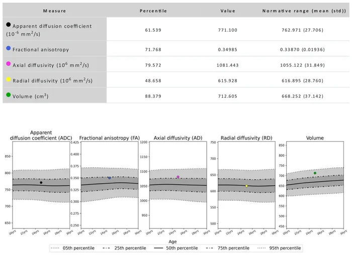
Measured against the normative range
Each bundle, and the white matter as a whole, is plotted for five measures (ADC, FA, AD, RD and volume) against healthy reference data, from the 5th to the 95th percentile.
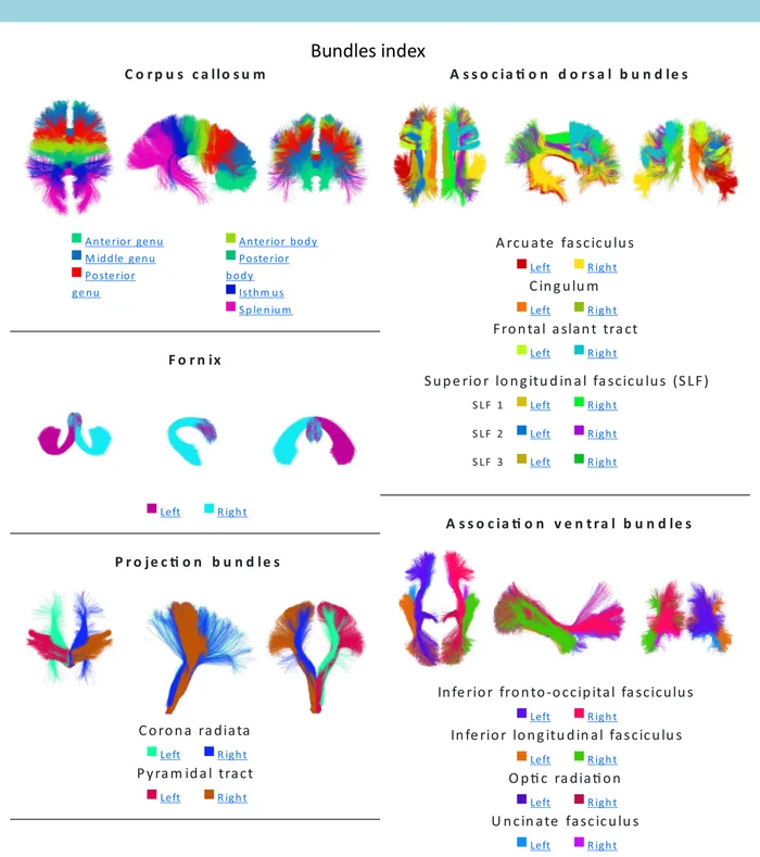
The 33-bundle atlas
Bundles are grouped as corpus callosum, association dorsal, fornix, projection and association ventral. This key belongs to the tract map, not to our brain illustration.
One layer of the read. Never the whole read.
Advanced DTI adds statistical evidence to a full neuroradiology read. It does not replace the conventional sequences, and it is not read from a picture.
-
Conventional MRI
T1, T2 and FLAIR images for structure and signal abnormality.
-
Susceptibility-weighted imaging
SWI to look for residual blood products.
-
Diffusion tensor analysis
Each tract measured and compared with healthy reference data by IMEKA’s ANDI.
-
The neuroradiologist’s impression
One signed report that weighs every sequence together.
A flagged tract is
- a measurement outside the normative range for the patient’s age and sex
- printed with its percentile, so anyone reading the report can check it
- read in context with every other sequence in the study
A flagged tract is not, on its own
- a diagnosis
- proof of a specific injury or its cause
- something to judge from left and right color differences on a tract map
Not every study comes back abnormal. When one does, the report names the tracts, gives the numbers and recommends clinical correlation.
Standard DTI and Advanced DTI, side by side.
| What’s in the report | Standard DTI | Advanced DTI |
|---|---|---|
| Radiologist’s brain read | Included | Included |
| Brain illustration | Included | Included |
| Simple patient explanation | Included | Included |
| Tract-by-tract analysis (up to 33 tracts) | Not included | Included |
| Full IMEKA/ANDI tractography report | Not included | Included |
| Plain-language summary of tractography findings | Not included | Included |
How each report is made
- 01 IMEKA/ANDI runs the tractography analysis.
- 02 We build a preliminary report from it.
- 03 A neuroradiologist reviews and signs it.
One report, read the same way for everyone who opens it.
- Imaging centers
Same scanners, a more detailed report.
Once your protocol is approved, your technologists send the study and we handle the analysis, the read and the report.
- Referring physicians
Findings tied to a specific tract.
Each flag comes with the measurement behind it, in a report a neuroradiologist signs.
- Legal teams
Documented tract by tract.
The normative comparison is printed in the report, beside illustrated pages written for laypeople.
- Patients
A summary in plain language.
It helps them understand their own imaging, alongside the radiologist’s report.
A brain injury report documents what the structural imaging demonstrates. It does not establish legal causation, liability, or case valuation.
Before you order one.
Who does the analysis?
IMEKA’s ANDI software runs the tractography analysis. We build a preliminary report from it, and a board-certified neuroradiologist reviews and signs every report.
Is ANDI FDA cleared?
Yes. ANDI received FDA 510(k) clearance in July 2023 (K230913), and ANDI 2.0 in October 2025 (K252298). The clearance covers measuring white-matter tracts as adjunctive information. ANDI does not make a diagnosis; the neuroradiologist’s report does.
How is Advanced DTI different from standard DTI?
Standard DTI is the brain read with an illustration and a simple patient explanation. Advanced DTI adds the tract-by-tract analysis, the full IMEKA/ANDI report and a plain-language summary of the tractography findings.
Does every study come back abnormal?
No. Each tract is compared statistically against healthy reference data, and the report states what it found either way, including when every tract falls within its range.
Why does the report have two color keys?
IMEKA/ANDI’s legend goes with the tract map. Our illustration key goes with the brain illustration and the patient summary. We keep them separate so each stays easy to read, and each applies only to its own section of the report.
How do I see a sample report?
Schedule a demo, and we’ll walk you through an anonymized Advanced DTI report.
See it for yourself.
Schedule a demo, and we’ll walk you through an anonymized Advanced DTI report, end to end.
Schedule a demo